Researchers at Stanford University have discovered a previously unknown immune cell in regenerative flatworms that destroys nearby threats by exploding within minutes. The study, published in Cell on June 2, 2026, identifies the cells as “ruptoblasts” and describes a rapid self-destruct process called ruptosis.
The finding came from planarian flatworms, tiny animals famous for rebuilding lost body parts. In experiments that fused tissue from different worms, the researchers saw an intense inflammatory reaction. Certain cells burst open, released toxic substances, killed nearby cells and then disappeared.
“We never expected that a cell could just explode like a bomb and kill the cells surrounding it,” said Bo Wang, senior author of the study and an associate professor of bioengineering in Stanford’s schools of Engineering and Medicine.
The discovery widens the map of immune biology. Most familiar immune defenses come from mammals, especially humans and laboratory mice. Planarians belong to a far older branch of animal life and their strange strategy suggests that nature has tried many more ways to fight infection and tissue invasion than scientists have fully cataloged.
Flatworms reveal ruptoblasts
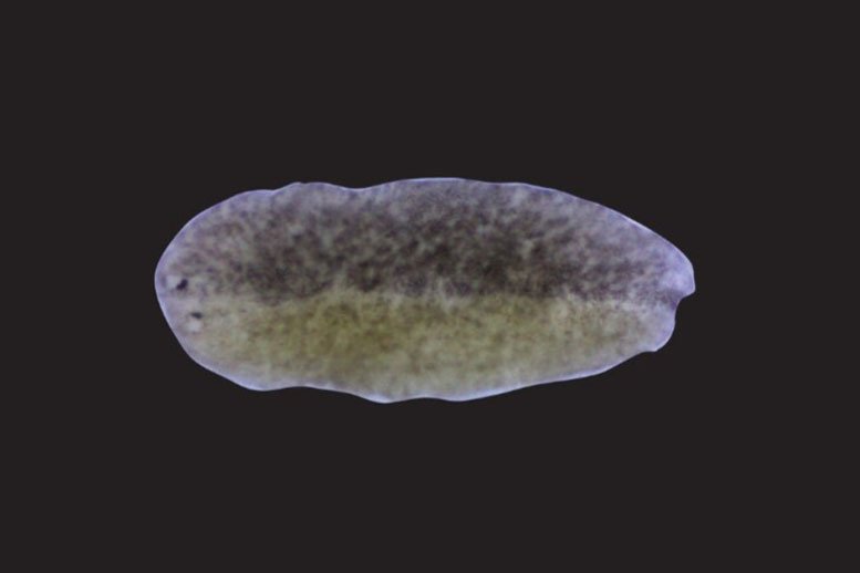
Planarian flatworms are small, soft-bodied animals with a remarkable ability to regenerate. Cut one into pieces and each fragment can rebuild missing tissues. That healing power has made them a favorite organism for scientists who study stem cells, wound repair and the rules that guide bodies back into shape.
In the new study, Stanford researchers found that planarians also carry a surprising immune weapon. The newly named ruptoblasts are cytotoxic cells, meaning they can kill other cells. Their method is unusually dramatic. When activated, a ruptoblast ruptures, releases harmful contents and destroys nearby targets.
The cells appear to be glandular, which sets them apart from the immune cells most readers know by name. T cells, natural killer cells and neutrophils are part of blood-forming immune lineages in vertebrates. Ruptoblasts point to a different path, one that links secretory cell biology with defense.
For a flatworm, that may be useful. These animals live in microbe-rich environments where bacteria and viruses are constant pressures. A fast local blast could help contain a threat before it spreads through soft tissue.
How chimera worms exposed the defense
The discovery began with a question about identity. Chew Chai, a postdoctoral researcher in Wang’s lab and lead author of the paper, wanted to know whether flatworms can distinguish their own tissues from tissues belonging to another individual.
To test that idea, she sliced worms lengthwise and fused them with tissue from separate worms. These blended animals are sometimes described as chimera worms because they contain material from different individuals. Planarians can regenerate their own missing parts with ease, but the researchers found that tissue from unrelated worms could be rejected.
That rejection resembled a broad biological alarm. Chai observed a strong inflammatory reaction in the fused animals. “It’s this huge inflammatory response,” she said.
The response showed that planarians have a way to detect biological mismatch. In humans, transplant rejection involves many well-studied immune players. In flatworms, the Stanford team saw a defense that led them toward a previously unknown cell type.
That path from tissue fusion to immune discovery is important. The researchers weren’t only watching flatworms heal. They were watching how a regenerative animal decides which tissue belongs and which tissue should be removed.
Activin sets off the blast
A key signal in the study was activin, a hormone already known to influence flatworm biology. Earlier work had shown that activin levels affect regeneration and reproduction. High levels can reduce the animal’s ability to regenerate, while low levels can interfere with reproduction.
When Chai studied the rejecting chimera worms, she saw activin rise along with chronic inflammation. The worms did not die immediately, but they often died within days. Similar inflammation appeared when healthy, unfused flatworms were injected with activin.
That result suggested activin was acting as more than a developmental or reproductive signal. In this context, it behaved like an inflammatory trigger. It helped connect tissue surveillance with immune defense.
To find the cells responding to that signal, the team used live-cell microscopy and flow cytometry. Live-cell microscopy let the researchers watch cells in action. Flow cytometry allowed them to sort stained cells one by one with laser-based analysis.
After labeling cells with fluorescent dyes, Chai isolated the population that reacted to activin exposure. Those cells were the ruptoblasts. Once activated, they released destructive material and vanished in a matter of minutes.
A self-destruct attack in minutes
The Stanford team named the explosive process ruptosis. It is a form of cell death that also functions as an attack. The cell sacrifices itself and the released contents kill targets in the immediate area.
Speed is one of the most striking features. “Ruptosis happens within seconds to minutes,” said Chai. That timing separates the process from slower forms of explosive cell death that have been described in some mammalian cells and bacteria.
The study found that ruptoblasts could damage several types of targets. The researchers tested them against E. coli bacteria, human kidney cells and mouse blood cells. The ruptoblasts killed all three in experiments, showing broad cytotoxic activity.
The damage stayed local. Cell death was limited to the area near the rupture, with no runaway chain reaction described in the Stanford account. That narrow range matters because any useful immune attack must balance force with control.
Inside the cell, ruptosis appears to rely on a sudden calcium surge from the endoplasmic reticulum, a structure that helps cells manage proteins and internal signaling. The study also points to the cytoskeleton, the cell’s internal scaffolding, as part of the signal amplification that drives the blast.
Why an ancient immune trick matters
Ruptoblast-like cells were found only in basal bilaterians when Chai looked across other animals. That pattern suggests an ancient evolutionary origin. It also raises a larger question about how different animal branches solved the problem of defense.
Wang sees that diversity as a reason to look beyond the usual laboratory organisms. “There’s lots of different immune mechanisms out there,” he said.
The flatworm strategy may reflect the animal’s unusual biology. A self-destructing immune cell can damage surrounding tissue and planarians have abundant stem cells that help them repair injuries. A vertebrate body with less sweeping regenerative power may have faced different tradeoffs during evolution.
The findings could also inspire biomedical thinking, although the work remains basic research in flatworms. Wang suggested that a highly localized destructive mechanism could offer ideas for future approaches to bacterial infections or tumors. Any medical application would require much more study, especially because controlled tissue damage is a demanding therapeutic goal.
For now, the discovery gives scientists a vivid new example of immune creativity. A tiny flatworm, best known for regeneration, has revealed a cell that turns self-destruction into defense. It’s a reminder that even familiar ideas such as immunity can look very different in the wider animal kingdom.