A study in PLOS ONE analyzed blood samples and MRI scans from 2,044 older adults in Japan and found that lower vitamin C levels were associated with smaller brain volumes and weaker connectivity in a major cognitive network. The research was led by Haruka Nagaya of Hirosaki University, with coauthors from Hirosaki University, Kyoto Prefectural University of Medicine and KAGOME CO., LTD.
The finding points to a measurable relationship between vitamin C levels in the blood and the physical structure of the aging brain. Participants with lower plasma vitamin C tended to show less gray matter and reduced connectivity within the default mode network. This network supports memory, attention, self-reflection and other internally focused mental processes.
Vitamin C is widely known for its role in immune health. In the brain, it also acts as an antioxidant and participates in processes that help nerve cells function. The new work adds brain imaging evidence to a broader question in aging science: how everyday nutrition may relate to the brain’s ability to maintain structure over time.
The researchers were careful about the interpretation. The study was cross-sectional, which means it captured a single window in time. The results show an association between lower plasma vitamin C and differences in brain structure. They do support a new hypothesis about nutrition and brain aging that researchers can test in longer studies.
Lower Vitamin C Was Tied to Less Gray Matter
Gray matter contains many of the brain’s nerve cell bodies and is central to information processing. In the PLOS ONE study, lower plasma vitamin C was significantly associated with a lower gray matter volume relative to total intracranial volume. That adjustment helped account for differences in head size across participants.
The analysis also found a significant association with white matter volume. White matter carries signals between brain areas through long nerve fibers. Together, the volume findings suggest that vitamin C status may track with broad features of brain structure in later life.
Researchers adjusted their analyses for many factors that can influence brain health. These included age, sex, education level, Mini-Mental State Examination score, diabetes, hypertension, hyperlipidemia, smoking history, drinking history and physical activity. Even after those adjustments, plasma vitamin C remained linked with gray matter volume.
The authors wrote that “plasma vitamin C levels are positively associated with the structural integrity of the gray matter and DMN connectivity.” In practical terms, people with higher measured vitamin C levels tended to have brain imaging patterns that looked more preserved within the areas the team examined.
That finding fits with vitamin C’s biological role as an antioxidant. The brain uses large amounts of oxygen and is vulnerable to oxidative stress. Vitamin C can help neutralize reactive molecules that may damage cells, which gives researchers a plausible pathway to explore.
A Key Brain Network Showed Weaker Connectivity
The study went beyond simple brain volume measurements. The researchers also examined structural connectivity within the default mode network, often called the DMN. This network includes several interconnected regions that are active during inward-focused thought.
Using MRI-based analysis, the team identified three gray matter structural networks related to the DMN. These were described as an anterior DMN, posterior DMN-I and posterior DMN-II. The posterior networks included regions such as the posterior cingulate cortex and precuneus.
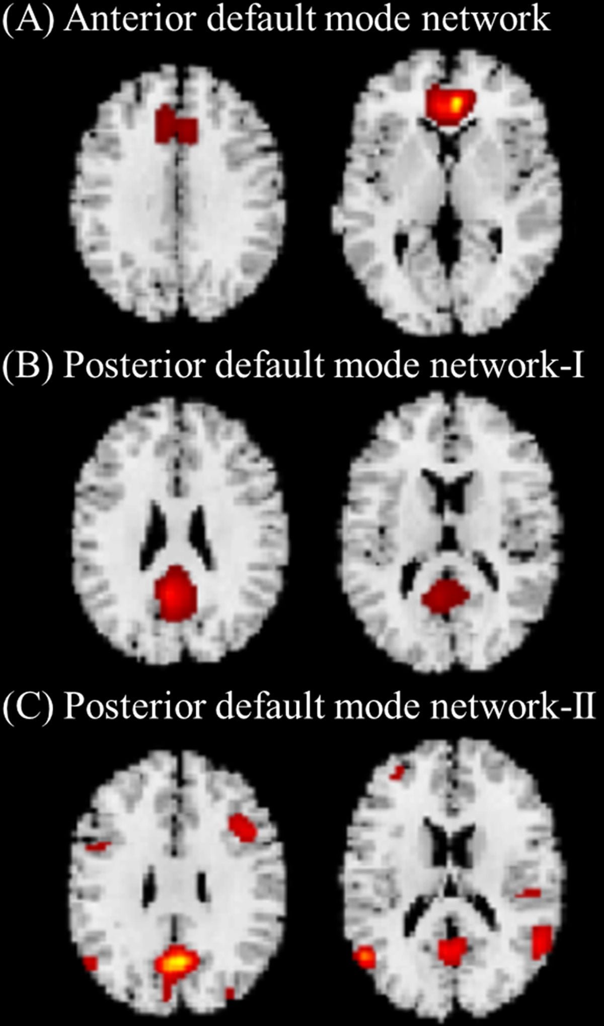
Participants with lower plasma vitamin C tended to have weaker connectivity within these DMN-related structural networks. The association remained significant after the researchers accounted for demographic, health and lifestyle factors. That makes the DMN finding one of the study’s most striking results.
Connectivity in this context refers to coordinated structural patterns across brain regions. The study used source-based morphometry, a method that looks for shared patterns in gray matter across the brain. For a large community-based cohort, this kind of MRI approach can reveal network-level differences that may be missed by simpler measurements.
Several regions highlighted in the analysis overlap with areas involved in memory and attention. The voxel-wise analysis found positive associations between vitamin C levels and regional gray matter in the posterior cingulate cortex, middle cingulate cortex, medial prefrontal cortex and inferior temporal regions.
What MRI Scans Revealed in 2,044 Older Adults
The study included 2,044 Japanese adults aged 64 and older. The participants had a median age of 69 years and 61.1% were female. Each participant underwent 3T magnetic resonance imaging, a high-resolution scanning approach commonly used to study brain structure.
Blood samples were collected after an overnight fast. The researchers measured plasma vitamin C, also known as ascorbic acid and compared those levels with brain MRI measurements. The team calculated total intracranial volume, gray matter volume and white matter volume.
For the network analysis, the researchers used independent component analysis to identify gray matter structural networks. Out of 147 gray matter structural networks, three were identified as related to the DMN. Those components then became the focus of the connectivity analysis.
This design gave the researchers two complementary views of the aging brain. One view measured overall tissue volume. The other examined large-scale brain networks that support cognition. Both pointed toward a relationship between plasma vitamin C and brain structure.
The sample size matters. A cohort of more than 2,000 people gives researchers more statistical power to detect subtle associations. It also helps separate the vitamin C signal from other influences such as diabetes, smoking, alcohol use and physical activity.
Why the Default Mode Network Matters
The default mode network is one of the brain’s most studied systems. It is active during resting or task-independent mental states. Scientists often link it to autobiographical memory, future thinking, attention, self-reference and understanding the thoughts of others.
In aging and neurological research, the DMN attracts attention because it includes brain regions that are vulnerable in several forms of cognitive decline. The posterior cingulate cortex and precuneus are especially important nodes. They help coordinate information across distributed brain areas.
The PLOS ONE study found that higher vitamin C levels were associated with better-preserved structural patterns in DMN-related networks. The anterior DMN and posterior DMN-I also showed positive correlations with Mini-Mental State Examination scores. That cognitive screening measure is widely used in older adults.
The researchers did find that plasma vitamin C levels were directly tied to brain structure more clearly than to MMSE scores. That distinction is important. Brain imaging may capture subtle changes before they appear as clear differences on a broad cognitive screening test.
For readers, the main takeaway is straightforward. The study connects a common nutritional marker with a brain network that supports everyday mental functions. It gives scientists a reason to ask whether maintaining healthy vitamin C status could support brain aging.
The Study Shows a Link, Not Proof of Cause
The results should be read with scientific caution. This was an observational study that measured vitamin C and brain structure at one time point. It can identify associations. It cannot prove that higher vitamin C levels directly caused better-preserved gray matter or DMN connectivity.
Dietary patterns are complex. People with higher vitamin C levels may also differ in other ways that matter for the brain. They may eat more fruits and vegetables, exercise differently, have different medical histories, or follow health behaviors that are hard to measure completely.
The researchers adjusted for several major health and lifestyle factors. Those adjustments strengthen the analysis, but hidden influences can remain in any observational study. Long-term studies are needed to see whether changes in vitamin C status come before changes in brain structure.
The study also focused on older adults in Japan. That makes the community-based cohort valuable and well defined. It also means future research should examine other populations with different diets, genetics, medical backgrounds and socioeconomic conditions.
For now, the findings add to a growing body of evidence linking nutrition with brain health. They also show how modern MRI methods can detect subtle network-level differences in large groups of older adults.
What Researchers Want to Test Next
Future studies can track vitamin C levels over years instead of measuring them once. That would help clarify whether stable vitamin C status predicts slower structural change in the brain. It could also show whether declining vitamin C levels appear before measurable brain changes.
Researchers may also need to examine diet in more detail. Plasma vitamin C reflects recent intake and the body’s handling of the nutrient. Broader dietary patterns, supplement use, fruit and vegetable intake and other antioxidants could all shape the relationship.
Mechanistic studies could explore how vitamin C might influence the brain. Possible pathways include antioxidant protection, support for nerve cell metabolism and effects on blood vessels that supply brain tissue. These possibilities need direct testing before they can guide clinical recommendations.
Intervention studies would be especially informative. Carefully designed trials could test whether improving vitamin C status changes brain imaging measures or cognitive outcomes in older adults. Such trials would need to consider baseline vitamin C levels, diet, health conditions and safe dosing.
The new findings make a focused case for further work. In a large sample of older adults, lower vitamin C in blood plasma was tied to less gray matter and weaker connectivity in the default mode network. That link gives brain aging researchers a clear target for the next wave of nutrition and MRI studies.