Researchers at Nagoya University have identified an unexpected self-modifying behavior in a brain-related enzyme, according to a study in the Journal of Biological Chemistry. The enzyme, called ST8Sia5L, can build a long sugar chain on itself. That chain then helps control whether the enzyme stays quiet, leaves the cell and later becomes active again.
The finding centers on polysialic acid, a long chain of sugar molecules that helps shape communication in the brain. These chains are strongly linked to brain development, learning, memory and neural flexibility. Scientists had long associated their production mainly with two enzymes, ST8Sia2 and ST8Sia4.
The new work adds a surprising third player. ST8Sia5 was already known as an enzyme involved in building fatty brain molecules called gangliosides. The long form, ST8Sia5L, now appears to have a hidden second ability. It makes polysialic acid on itself, then uses that modification as part of a built-in control system.
A Hidden Ability in ST8Sia5L
ST8Sia5L belongs to a family of enzymes that attach sialic acid, a type of sugar building block, to other molecules. These enzymes help make specialized glycans, which coat cells and influence how they behave. In the nervous system, those surface sugars can affect how strongly cells attach, how signals are presented and how flexible neural circuits remain.
The research team found that ST8Sia5L can create polysialic acid through autopolysialylation. In plain terms, the enzyme decorates itself with a long sugar chain. The study discussion describes the result this way: “ST8Sia5L is a new polysialyltransferase that autopolysialylates its N-glycans.”
That matters because ST8Sia5 had been known for a different task. It helps modify gangliosides, fatty molecules that sit in cell membranes and are abundant in the brain. Gangliosides influence how neurons communicate and how receptors are organized on cell surfaces.
The new activity appeared only in the long form of the enzyme. ST8Sia5 exists as short, medium and long forms. The study indicates that the long version occupies a different internal cell compartment than the shorter versions. That location may help explain why ST8Sia5L can perform this unusual self-modification.
The Brain Sugar Chain Behind the Discovery
Polysialic acid is a long chain made from repeating sialic acid units. In the brain, it is best known for modifying the neural cell adhesion molecule, often called NCAM. When NCAM carries polysialic acid, brain cells can adjust how tightly they stick to one another.
This slippery effect is valuable during development, when neurons migrate and form new connections. It also remains important in selected regions of the adult brain that continue to remodel. For that reason, polysialic acid is often associated with neural plasticity, the brain’s ability to adapt.
Polysialic acid also binds growth factors and neurotrophins. These are signaling molecules that help neurons survive, mature and respond to changing conditions. By holding or presenting these molecules near cell surfaces, polysialic acid can influence how signals reach their receptors.
The study adds ST8Sia5L to the short list of enzymes capable of building long polysialic acid chains. The distinction is precise. ST8Sia5L builds the chain on itself, while ST8Sia2 and ST8Sia4 are mainly known for adding polysialic acid to other molecules such as NCAM.
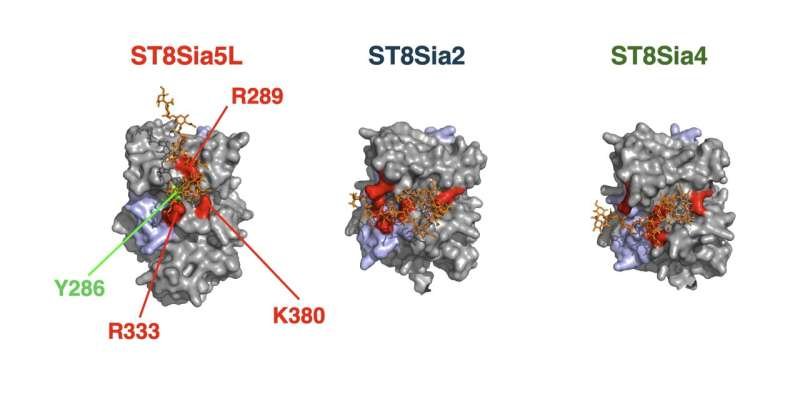
The image compares three polysialic acid-building enzymes. ST8Sia5L is shown with amino acids that help its polysialic acid synthesis. Image credit: Sakamoto et al., 2026.
How the Enzyme Turns Itself Off
The self-built sugar chain appears to act as a molecular switch. When ST8Sia5L is coated with polysialic acid, its ganglioside-building activity is suppressed. After the chain is removed, the enzyme can regain that activity.
This gives the enzyme a compact control system. It carries out one chemical job on itself, then that modification changes its ability to do another job. The study found that secreted, autopolysialylated ST8Sia5L showed no ganglioside-sialylation activity. Treatment that removed polysialic acid restored the enzyme’s activity in experiments.
The researchers also identified structural clues behind this behavior. Their analyses pointed to a polysialic acid trapping motif in ST8Sia5L. This motif appears to support the elongation of the polysialic acid chain. It differs from the conventional domain associated with ST8Sia2 and ST8Sia4.
Several amino acids appear important for the process. The study highlights residues including R289, R333, K380 and Y286 in ST8Sia5L. These sites help explain how the long form can build polysialic acid in a way the shorter forms do not.
Why Secretion Matters
Once ST8Sia5L has modified itself with polysialic acid, the enzyme can be released from the cell. The study links this release to metalloprotease enzymes, which can cut proteins near membranes. In experiments, inhibitor tests pointed to metalloprotease involvement rather than exosome-based release.
This secretion step makes the discovery more intriguing. Enzymes that build glycans are commonly discussed in the context of intracellular processing. Cells usually assemble many sugar modifications inside compartments such as the endoplasmic reticulum and Golgi apparatus. The ST8Sia5L result suggests a route that could place an active enzyme outside the cell after its self-built chain is removed.
The team also found that ST8Sia2 and ST8Sia4 can be secreted in polysialic acid-coated forms. Their functions after secretion remain unclear. Still, the observation broadens the question beyond one enzyme and raises the possibility that secreted polysialylated enzymes may have underappreciated roles.
For ST8Sia5L, secretion and silencing seem to be connected. The sugar chain is associated with the enzyme leaving the cell and the same chain keeps its ganglioside-building function quiet. Removal of the chain changes that state.
A Possible Fast Repair System for Brain Lipids
Gangliosides are a major part of neuronal membranes. They help organize surface molecules, support signaling and shape how neurons respond to their environment. Because these molecules sit at the cell surface, changes to them can affect communication between brain cells.
The Nagoya University team proposes that secreted ST8Sia5L may help repair or restore ganglioside structures directly at cell surfaces. This remains a hypothesis. The study’s cell-based experiments show that the enzyme can be secreted and reactivated after polysialic acid removal. The next question is whether that same sequence occurs in living tissue.
A fast, local repair pathway would be biologically useful. If a ganglioside structure is damaged or altered at the surface, a nearby enzyme could help restore it without requiring the molecule to travel deep into the cell. The researchers are investigating this proposed on-site recovery mechanism.
The idea also fits with the reversible nature of the enzyme’s control system. ST8Sia5L can leave the cell in a quiet state while coated in polysialic acid. Once the coat is removed, the enzyme could resume ganglioside-related activity in the extracellular space.
Links to Inflammation and Brain Disorders
The study also points toward possible connections with stress, inflammation and brain disease. During stress or immune activation, cells can release sialidase enzymes. These enzymes remove sialic acid-containing structures, including polysialic acid.
If sialidases remove the polysialic acid coat from secreted ST8Sia5L, the enzyme could regain activity outside the cell. That would link inflammation-related sugar removal with ganglioside modification. The study presents this as a possible mechanism to explore, rather than a proven disease pathway.
Another question involves microglia, the immune cells of the brain. The researchers hypothesize that polysialic acid-coated ST8Sia5L may interact with inhibitory receptor molecules called Siglecs. Such interactions could help restrain immune activation under normal conditions.
Polysialic acid abnormalities have also been associated with schizophrenia. The mechanism behind that association remains unresolved. The secreted polysialylated form of ST8Sia5L is now a candidate for further study in that context, especially because it combines polysialic acid biology, enzyme regulation and brain-specific lipid modification.
What the Researchers Will Test Next
The current findings come from biochemical and cell-based experiments. That makes the mechanism strong at the molecular level, while leaving important biological questions open. The next step is to test whether ST8Sia5L behaves the same way in living organisms.
To address that, the team is generating mice in which the ST8Sia5 gene has been disabled. Such animals could help show whether the enzyme affects brain development, neural repair, immune regulation, or behavior. They may also reveal whether other enzymes compensate when ST8Sia5 is missing.
The shorter forms of ST8Sia5 remain another open area. ST8Sia5S and ST8Sia5M localize differently inside cells and their functions are still unclear. Comparing these forms with ST8Sia5L could show how small structural changes redirect an enzyme’s location and activity.
The work also gives glycobiology a broader question to pursue. Sugar chains often act as signals, shields and binding surfaces. In ST8Sia5L, a sugar chain becomes part of an enzyme’s own control circuit. That simple twist opens a new view of how brain cells may regulate molecular tools at their surfaces.